Aidan's Hypophosphatasia Friends
Aidan's friends are children and adults just like him. Friends who are fighting Hypophosphatasia. Friends who hurt, but don't know why. Friends who know a doctor and dentist office only to well. Friends trying to figure out what is happening to their bodies. Why their teeth keep falling out, or their bones keep breaking. This page is for those friends. You are not alone.
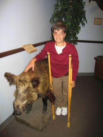 Sue comes to us from the SW. She is 53 years old and was diagnosed within days of birth. It was also discovered she had fractured bones while in the womb. Sue's mother says when born, Sue looked like a rag doll with no strengthens at all, not even to grasp a finger. Sue had blue sclera, a large fontanel, low APL, bowed long bones, and short arms. Her first bi-lateral displaced femur fractures were at three months old trying to roll in bed. While in the ER they took a whole body x-ray and found that Sue had other fractures in different healing stages. As an infant she fought weight gain until someone suggested a meat based formula. She continued to fracture all upper and lower long bones at least two times a year. In the fall of 1965, Sue was on her 14th double displaced femur fractures, total broken bones including finger, toes, wrist, and ribs compression fractures of the vertebrae 26. It was after much testing six different doctors determined Sue had Osteogenesis Imperfecta and not Hypophosphatasia. Her APL levels were low but her B and calcium levels were normal. Back then treatment was basically the same. In 1966 Sue went to see Dr. Sofield at Shriner's. In 1967 Sue had her first rodding surgery of her femurs. In six months she had the other femur done and then the tibia. By 1968 Sue was able to walk around with crutches. She was never able to do this before the bone roddings; the bones were too weak and would fracture without any trauma. The re-rodding surgeries happened about every one to two years because Sue was growing and in that amount of time the bones would start to bow again and the fractures would happen in the unprotected portion of the long bone. This went on until Sue was 14. At this point she had to get a new orthopedic doctor (her original doctor retired) and her new doctor felt she had some form of HPP; he just wasn't sure what form. In 1978 Sue's femoral rods needed to be replaced. Since she moved from home for college, a new orthopedic doctor was needed again. He diagnosed her with OI, with unexplained very low APL and high normal B6 with low normal calcium so he said she couldn't be HPP. She was labeled OI with unexplained TNSALP. Sue stayed OI until 2009 when her primary care doctor wanted her to see another endo doctor, her APL was down to 3, and he told her you couldn't even say you had any APL in your body. In the previous five years Sue had 4 new re-roddings of the long bones, cervical neck fusion with hardware (front plate 8 screws and 2 rods in the back) from all the micro fractures and a compression fracture in her vertebrae causing Kyphosis and spinal cord compression. She also had 1 total hip replacement with long stem in her right hip due to protrusio. Since both HPP and OI are so rare, hardly any doctors fully know the story about either metabolic disease. The tests came back wit. h mixed results, 50% leaning toward HPP while 50% leaned toward OI. The doctor couldn't find a lab in the USA that ran test for Phosphoethanolaminuria (PEA) so he couldn't say 100% that Sue had HPP over OI. The doctor said Sue could have both; it's possible, very very rare but possible. Sue had a DNA test for OI, which isn't reliable; it has a 70% false negative result for type 4 OI which is Sue's clinical diagnosis. DNA testing for OI just started in 2005, so it was possible to get negative results, but it didn't mean Sue could rule it out. It simply meant they did not have a classification yet for Sue's DNA mutation. In March 2010 Sue's DNA test came back showing 90% that she does not have OI. Sue will be having blood drawn by her endo doctor that will sent to Dr. Whyte in St. Louis for DNA testing for Hypophosphatasia. Sue has continued to have at least 3 fractures not all severe every year of her life. 2009 was Sue's biggest year of spontaneous fractures in the last 11 years. Sue had two dislocated shoulders just getting dressed with hairline fractures of her humerus, two compression fractures of the thoracic vertebra and a cracked ankle and wrist just doing every day stuff like getting dressed, making the bed, etc. Sue has had over 60 fractures and multiple roddings. Since last December Sue broke a wrist, elbow, shoulder, fibula, ankle and 3 ribs.
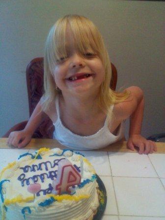 Corinna comes to us from Pennsylvania and is 6 years old. Lauren, Corinna's mom, shares Corinna's story with us. Corinna (CC) was diagnosed with childhood type Hypophosphatasia when she was around 6 months old. It was kind of a fluke how we found out. When she was 6 weeks old my son was playing with her and broke her arm. I took her to the Doctor who sent me to the emergency room. When she got her x-ray, the Dr. noticed something looked funny, so he called her Dr. and ordered a full set of x-rays. That is when they saw "holes" in her growth plates. She was admitted and stayed for 4 days. Then they diagnosed her with cartilage hair Hypophosphatasia, a rare form of dwarfism. I searched the Philadelphia area for a Dr. that specialized in dwarfism, I found Dr. Michael Bober in the genetics department at DuPont Hospital for Children in Delaware. I made an immediate appointment. As soon as Dr. Bober saw Corinna, he informed me that she did not have CHH but he thought she had a disease called Hypophosphatasia. Dr. Bober then sent her blood off to the lab to be genetically tested. She tested positive for HPP. So here we are 4 years later. The symptoms CC shows are knobby knees, she has about 6 teeth and she has scoliosis. Corinna also has some joint pain and her one leg is longer than the other. She is currently enrolled in the Enobia drug study and is doing great! Watch Corinna's amazing progress thanks to Enobia and the enzyme replacement therapy.
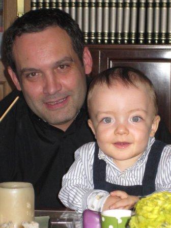 Arturo hails from Zaragoza, Spain. Cesar, Arturo's dad shares Arturo's story with us. Arturo was diagnosed in December 2010 when he was 17 months old. Arturo can not walk and is underweight. Vomiting has been constant. He is happy and brings happiness. After 8 months we have much hope with Enobia. Dad says, "I will do all I can to restore the happiness that I get."
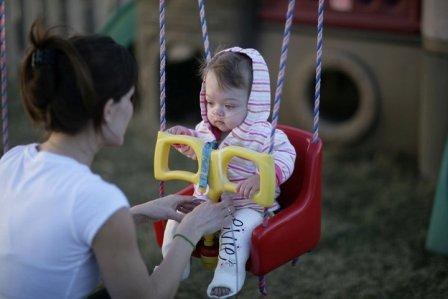 Evie is a bright eyed little girl from Nebraska, who turned four in September of 2013. When her mom was pregnant there was little hope that she would survive but she has beaten the odds and proved her doctors wrong. At two weeks of age Evie was diagnosed with severe infantile hypophosphatasia with seizures and was given a 50% chance of survival. Untreated her doctor anticipated she would survive 5 or 6 months. Evie started the Enobia trial at two months of age and has shown significant improvement. Her orthopedist has commented that her x-rays do not look like they could possibly be from the same child. She is growing new bone and her seizures seem to be requiring less medicine. Her parents' hope that someday she will walk on her own and continue to progress. Evie's first year was filled with appointments, hospitalizations and a few surgeries but her second year has been a blessing to her and her family. We are thrilled that she has done so well and pray for continued growth and support for the HPP community. Evie and HPP make the news. Evie's HPP story as published by Reuter's. Evie also has her own blog which mom updates regularly.
 Casey shares his story of HPP with us. I was born November 2, 1979. Everything looked good at birth. I suffered from asthma and bronchitis. My PCP was keeping track of the growth of my skull. One time during a regular visit, my doctor said "enough", my cranium growth was off the charts and I needed to go to the hospital. First, they thought I had rickets due to the nodules on my ribs. After several labs and scans, they still couldn't figure out what was wrong. A doctor consulted a medical book and put everything together and finally diagnosed me with HPP. I had craniosynostosis and all the sutures on my skull were closed and my brain was about to come out of my soft spot. They performed surgery and made their own sutures, which was a success. The only thing they told my parents about the lasting effects of Hypophosphatasia would be, crippling arthritis and that I would be in a wheelchair by the age of 25. During my childhood, I lost my bottom front teeth as soon as they came in. I was made fun of by other children due to my misshaped head. I could never run like other children as I was always kind of stiffed legged. Pain would set in after running or climbing stairs, etc. with the pain being in the shins, calves, and feet. I tried playing baseball, which didn't work out to well. After every game or practice, it was a hot bath and baby aspirin, which never did work well, but my parents didn't know what else to do. I tried playing for three seasons, never hit a ball during a game, but I had a good time and made quite a few friends. My adolescent years were uneventful; I didn't suffer much of anything. What pain I had dealt with through the previous years, I was accustom to.
Everything started in my mid twenties. Standing long periods of time would result in my feet, legs and lower back hurting. I suffered some hip pain and my hips would pop. My arms would tire quickly and my hands even faster. My left arm was, and is considerably weaker than the right. I kind of chalked this up to being out of shape or something. I experience pain in the back of my head that carries down the spine, in between the shoulder blades. My sternum started popping when I would stretch. Then at 26, after eating dinner one night, BAM, I had a grand mal seizure. I went to the hospital and found out I have Chiari Malformation 1 with a herniation of 9mm, syringoymyelia and scar tissue on the left temporal and frontal lobe. I suffer from three types of seizures; simple partial, complex partial and grand mal. Treatment has been successful. In four years I have had fifteen grand mal seizures and countless simple partial and complex partial. My bone problems seem to be getting worse. My shins hurt all the time like somebody took a stick and beat them over and over again, sometimes worse than others. My sternum pops more than ever when I reach or stretch. My right hand gets tired very easily, especially when writing. My left arm and left leg seem to be getting weaker, knees and elbows are starting to pop, which I never really experienced. Of course, all digits pop, when big toes pop pain sets in. I suffer floaters and orbs in my left eye. I am seeing a list of doctors, from primary care to sports medicine; none of them seem to know what to do. Osteoporosis? Osteopenia? I know for sure that my level is 9, calcium is high, INTACT PTH is low, and vitamin D is high. Below are the actual results of the bone axial density test and bone scan, but the doc said "I was good".
Clinical History: The patient is a 31 year old man with a history of bilateral leg pain.
Technique: The patient was injected intravenously with 25 mci of Tc 99m MDP. Routine delayed images of the entire skeleton were obtained.
Findings: The patient has an elongated skull which may represent craniosynostosis. There are small foci of increased tracer uptake at the costovertebral junctions of ribs 6 and 7 on the right. There is a small focus of increased tracer uptake at the first metatarsal-phalangeal joint of the left foot. There's no abnormal tracer activity associated with the femurs tibia or fibulae. Two kidneys are identified. Radioactive urine is seen in the bladder.
Comments: DXA of the lumbar spine (L2-L4) reveals a bone density of 0.981 g/cm2. This value is 1.2 standard deviations below the mean for young adults, and represents 88% of the mean density for patient's age. Femoral neck bone density is 0.769 g/cm2. This value is 1.2 standard deviations below the mean for young adults. Total hip density is 0.794 g/cm2, a value that represents 1.6 standard deviations below the mean for young adults. Hip density represents 78% of the expected density for patient's age. Density at the 1/3 radius is 0.657 g/cm2. This value is 3.0 standard deviations below the mean for young adults.
Summary: Osteoporosis in the 1/3 radius. Osteopenia in the lumbar spine, femoral neck and total hip. Patient has low z-score; consider secondary cause(s) of osteoporosis. Bone density at the 1/3 radius was below the expected range for age based on z scores which are used to define bone density in men less than 50 years of age. Bone densities at the lumbar spine, at the femoral neck and at the total hip were within the expected range for age based on z scores.